A pineal cyst is a type of brain cyst. A brain cyst is an abnormal fluid-filled sac in the brain. A cyst in the brain may contain cerebrospinal fluid (CSF). CSF normally bathes and cushions the brain and spinal cord. Often, a brain cyst starts before birth. They are generally not cancer (benign). Benign also means that the growth doesn't spread to other parts of the body. A pineal cyst can appear in people of any age.
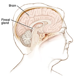
Understanding the pineal gland
The pineal gland is a small organ in the middle of the brain. It makes melatonin. This is the hormone that regulates sleep. A pineal cyst usually only shows up on an imaging scan done for another reason. A pineal cyst seldom causes problems. If it grows large, it can affect your vision.
What causes a pineal cyst?
Researchers are not sure what causes a pineal cyst.
Symptoms of a pineal cyst
A pineal cyst does not usually cause any symptoms. In rare cases, extra CSF or bleeding into the cyst may cause headache or make it hard to look up.
In rare cases, a pineal cyst may cause CSF to build up on the brain. This is called hydrocephalus. It's caused when the cyst blocks the flow of CSF in the brain. Hydrocephalus can cause symptoms such as:
- Confusion.
- Vertigo.
- Double vision.
- Headaches.
- Upset stomach (nausea).
- Sleepiness.
- Trouble walking.
- Vomiting.
- Passing out (syncope).
- Coma.
Diagnosing a pineal cyst
A pineal cyst is usually found by chance on an imaging test of the brain. This may be one of the following:
- CT scan. This is a test that uses a series of X-rays and a computer to create images of the inside of the body.
- MRI. This test uses large magnets and a computer to create images of the body. MRI scans of your brain may be done to get more information about the cyst and nearby tissues.
Treatment for a pineal cyst
A pineal cyst is usually only treated if it causes symptoms. If symptoms are present, your doctor may recommend these treatments:
- Endoscopic removal. This is surgery through a small tube called an endoscope.
- Stereotactic aspiration. This is the removal of fluid from the cyst with a needle. Imaging is used to help guide the needle.
Your doctor will talk with you about which treatment works best for you.
When to contact your doctor
Contact your doctor right away if you have any of these:
- Confusion
- Trouble walking
- Sudden headache
- Vision problems
Online Medical Reviewer: Anne Fetterman RN BSN
Online Medical Reviewer: Luc Jasmin MD
Online Medical Reviewer: Raymond Kent Turley BSN MSN RN
Date Last Reviewed: 12/01/2022
© 2000-2026 The StayWell Company, LLC. All rights reserved. This information is not intended as a substitute for professional medical care. Always follow your healthcare professional's instructions.Related Articles
Encephalitis
Encephalitis is a viral infection of the brain. It may cause headache, stiff neck, irritability, fever, drowsiness, nausea and vomiting.
Understanding Metabolic Encephalopathy
Metabolic encephalopathy is when the brain has trouble working because of a chemical, or metabolic, problem in the body. Knowing more about this health problem can help you make the best choices about the care you may need.
Brachial Plexus Injury
Learn about brachial plexus injury, including cause, risk factors, symptoms, diagnosis, and treatment.
Peroneal Nerve Palsy
Peroneal nerve palsy is weakness in the muscles that lift the foot. This is often due to pressure on the peroneal nerve, which can have a variety of causes.