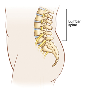
Fusing vertebrae in the lumbar spine may help ease lower back and leg pain. Spinal fusion is fusing two or more vertebrae together so they heal into a single, solid bone. The goal of spinal fusion is to reduce or eliminate painful motion and restore the stability of the spine. Posterior lumbar fusion is done through an incision in your back. Spacers and/or a bone graft are put between the vertebrae. Depending on how many vertebrae are fused, the surgery may take from 3 to 8 hours.
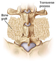
Fusing the transverse processes
Fusion has several steps:
-
The bone graft is packed between the transverse processes (“wings”) on the sides of the vertebrae. This procedure is called posterolateral fusion.
-
To help keep your spine steady and promote fusion, extra support may be used such as titanium screws and rods. Sometimes, bone cement is added around the screws.
-
A wound drain is often placed in the wound and left in for a few days.
-
The incision is closed with stitches (sutures) or staples.
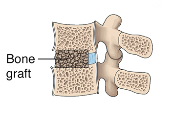
Fusing the disk space
This involves the following:
-
An incision is made in the middle of or on each side of your spine.
-
The disk between the vertebrae is removed. This procedure is known as posterior lumbar interbody fusion (PLIF).
-
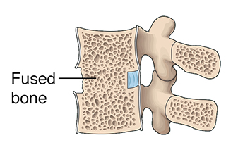
A bone graft, a spacer (cage), or both are placed in the now-empty disk space between the vertebrae. In time, the graft or spacer and the bone will grow into a solid unit.
-
To help keep your spine steady and promote fusion, metal screws, and rods may be placed to provide extra support and promote healing. Sometimes, bone cement is added around the screws.
-
A wound drain is often placed in the wound and left in for a few days.
-
The incision is then closed with stitches or staples.
Online Medical Reviewer: Anne Fetterman RN BSN
Online Medical Reviewer: Heather M Trevino BSN RNC
Online Medical Reviewer: Vinita Wadhawan Researcher
Date Last Reviewed: 03/01/2024
© 2000-2026 The StayWell Company, LLC. All rights reserved. This information is not intended as a substitute for professional medical care. Always follow your healthcare professional's instructions.

Related Articles
Understanding Clavicle Fracture Open Reduction and Internal Fixation (ORIF)
Open reduction and internal fixation (ORIF) is a type of treatment to fix a broken bone. It puts the pieces of a broken bone back together so they can heal.
Kneecap (Patella) Surgery: Realignment
Some procedures may be done using arthroscopy, a method that uses tiny incisions and special instruments to look and work inside the knee joint. Other procedures need open surgery. The kneecap can be realigned to improve its tracking. To do this, soft tissue may be cut, tightened, or moved.
Upper Arm Fracture Open Reduction and Internal Fixation (ORIF)
Open reduction and internal fixation (ORIF) is a type of surgery used to stabilize and heal a broken bone. You may need this procedure to treat your broken arm. Read on to learn what to expect before, during, and after this surgery.
Hip Arthroscopy: Repairing Femoroacetabular Impingement
When excess bone forms on the edge of the ball or the socket of the hip, it's called FAI (femoroacetabular impingement). This can cause pain and limit movement. Arthroscopy can fix FAI. It uses small incisions and special tools. Read on to learn more.